Kreuzbein (Os sacrum) Anatomie, Aufbau, Bänder & Gelenke Kenhub
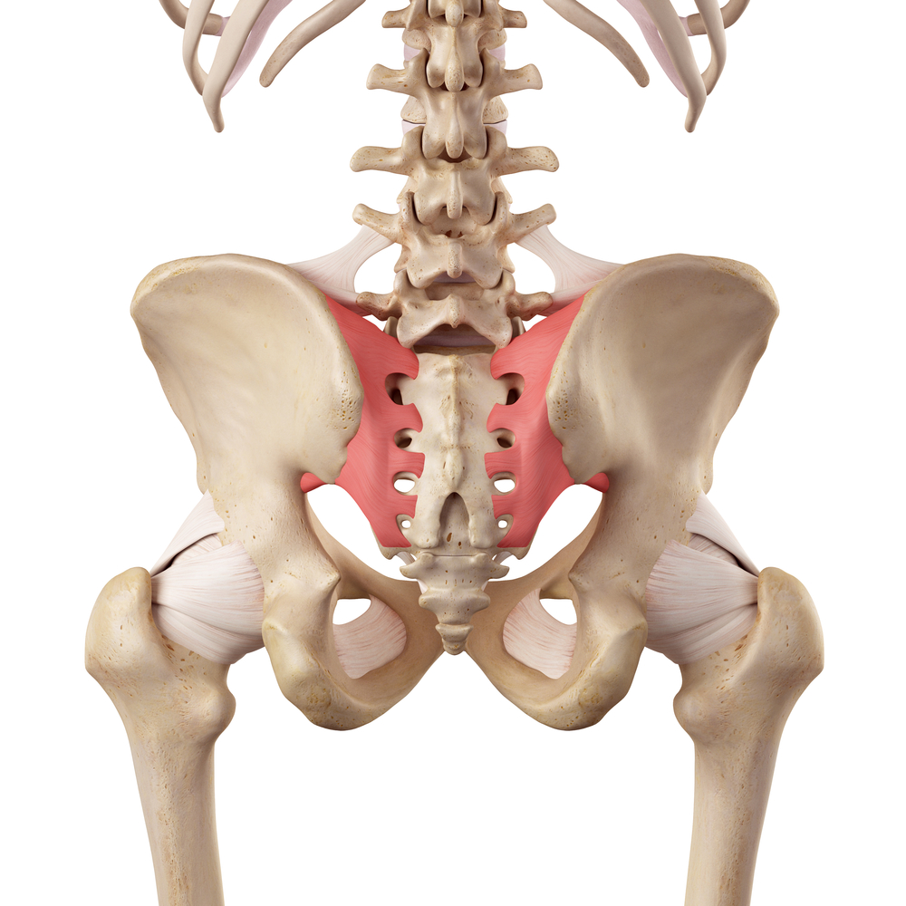
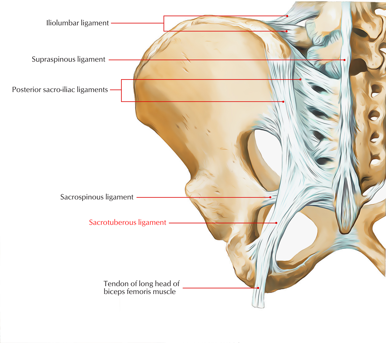
Die Ligamenta sacroiliaca anteriora überbrücken auf der Ventralseite den Gelenkspalt. Ligamenta sacroiliaca posteriora et interossea Die Ligamenta sacroiliaca posteriora und die Ligamenta sacroiliaca interossea ziehen von der Tuberositas iliaca zum Kreuzbein und verhindern, dass das Kreuzbein in die Beckenhöhle rutscht. Ligamentum iliolumbale
The Sacroiliac Joint
The anterior sacroiliac ligament consists of numerous thin bands, which connect the anterior surface of the lateral part of the sacrum to the margin of the auricular surface of the ilium and to the preauricular sulcus . See also Posterior sacroiliac ligament References

Figure 3 from [The pelvic girdle and the pelvis]. Semantic Scholar
Wikipedia

Sacrococcygeal Joint
beinbänder (Ligamenta sacroiliaca interossea), wie sie in der Fachliteratur immer wieder erwähnt werden, vorhanden sind. Weiterhin wird überlegt, ob im F alle der Ligamenta sacroilia-
Sacroiliac Joint Syndrome Physiopedia
TheFreeDictionary interosseous sacroiliac ligaments in·ter·os·se·ous sa·cro·il·i·ac lig·a·ments [TA] short obliquely directed fibrous bands that pass between the sacrum and ilium in the narrow cleft behind the auricular surfaces of these bones. Synonym (s): ligamentum sacroiliacum interosseum [TA] Farlex Partner Medical Dictionary © Farlex 2012

medical accurate illustration of the sacroiliac ligament Stock Photo Alamy
Die Ligamenta sacroiliaca interossea sind Teil des Bandapparates des Iliosakralgelenks. Anatomie Die kräftigen Bänder verlaufen zwischen der Tuberositas iliaca und der Tuberositas ossis sacri. Es handelt sich überwiegend um kurze, quer verlaufende Faserzüge. Funktion
Anterior interosseous artery Anatomy, branches, supply Kenhub
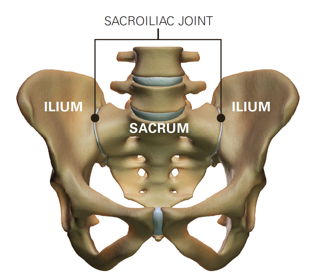
Das Iliosakralgelenk (Art. sacroiliaca, Kreuzbein-Darmbein-Gelenk) verbindet das Os ilium (Darmbein) und das Os sacrum (Kreuzbein). Charakteristisch ist der ausgeprägte Bandapparat, welcher die Beweglichkeit des Gelenks stark einschränkt. Articulatio sacroiliaca Kreuzbein-Darmbein-Gelenk 1/2
Sacroiliac Joint North Lakes Chiropractic
characteristics of the joint capsule: short, stiff, strengthened by these ligaments: anterior sacroiliac ligament posterior sacroiliac ligament sacroiliac interosseous ligament iliolumbar ligament
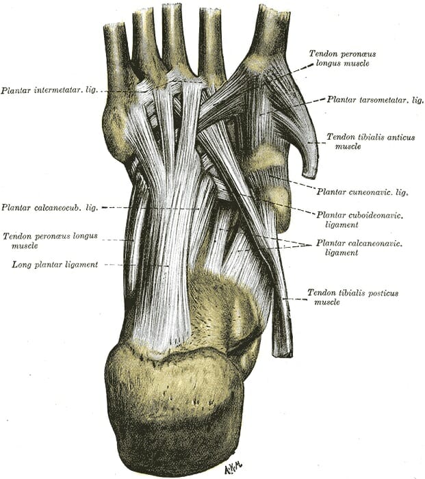
Foot (Anatomy) Bones, Ligaments, Muscles, Tendons, Arches and Skin
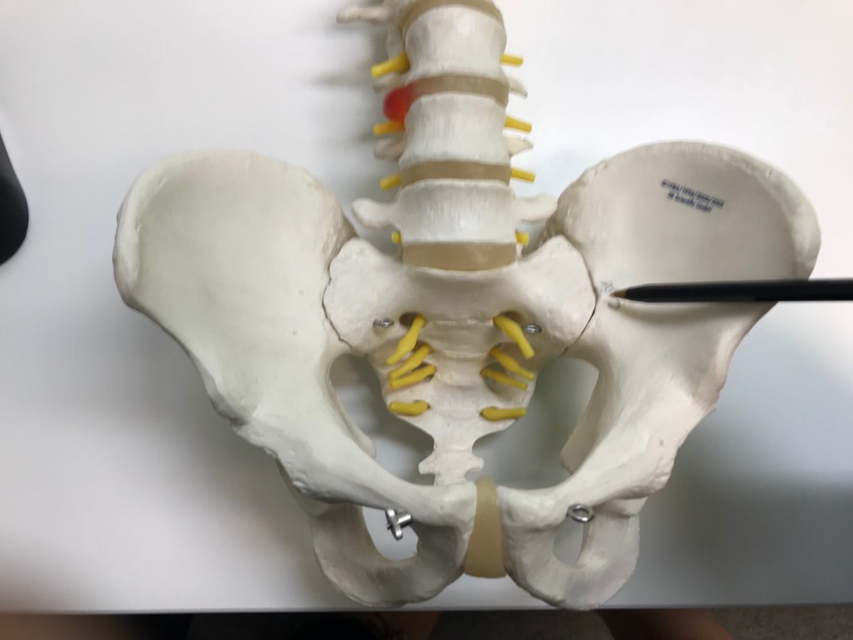
The sacroiliac joint is formed by the irregularly-shaped (jagged) articular surfaces of the sacrum and the ilium, which provide some strength to the joint 1,2. The articular surfaces of the ilium and sacrum are called the auricular surfaces due to their "L-shaped" resemblance to the pinna of the external ear. The upper one-third is a syndesmosis.

Pin on anatomy
These joints sit where the lower spine and pelvis meet. Sacroiliitis can cause pain and stiffness in the buttocks or lower back, and the pain might go down one or both legs. Standing or sitting for a long time or climbing stairs can make the pain worse. Sacroiliitis can be hard to diagnose. It can be mistaken for other causes of low back pain.

The SI Joint, Its Anatomy and Relationship to Yoga Practice
The sacroiliac joint has the strongest ligament system in the body. They are categorized according to their function of limiting nutation or counternutation. Ligaments limiting nutation include the sacrospinous, sacrotuberous, anterior capsule, anterior sacroiliac ligament, anterior longitudinal ligament, interosseous ligament, and the short posterior ligaments [1]p62-65 [2]p55-60 and the.
Sacrotuberous Ligament Earth's Lab
- dorsal sacroiliaca ligament (strong) - interosseal sacroliliaca ligament (strong) 2. Pubic symphysis - symphysial surface of pubis and an interpubic discus - synchondrosis, fibrocartilage Not a true joint, however, it may contain a „cavity" within the disc Ligaments: superior pubic lig. (inf.!) arcuate pubic lig.

6 Région sacroiliaque Medicine Key
The AIL failure loads are just slightly higher than those for the ligamentum flavum in the spine, a tissue composed mainly of elastic fibers. In contrast, the AIL has negligible elastin content. Because the AIL represents about 14% of the total area of interosseous sacroiliac ligaments, its mechanic.
Si Joint Pelvis and Hips
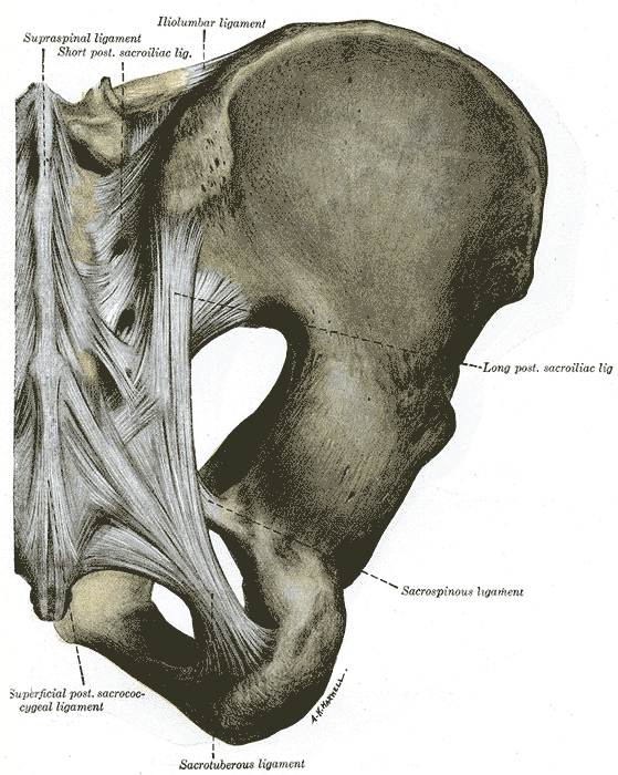
Definition Always much stronger than the ventral sacroiliac ligament, the dorsal sacroiliac ligament (Ligamenta sacroiliaca interossea) is subdividable into two parts, a short and a long one, that are often described as two distinct ligaments (Ligamenta sacroiliaca dorsalia) but between which it is difficult to make a demarcation.
/GettyImages-87394799-56a05fac5f9b58eba4b0275a.jpg)
Sacroiliac Joint Anatomy and Characteristics
Auxiliary facilities: discus articularis, ligamentum radioulnaris palmare and dorsale (articular disc, radioulnar palmar and dorsal ligament). sacroiliac ligaments), ligg.sacroiliaca interossea (interosseal sacroiliac ligaments) Type of joint: amphiartrosis Movements: minimal . 2. Symphysis pubica
Sacroiliac joint Anatomy, function Kenhub
Auxiliary facilities: lig. radiocarpeum dorsale and palmare (dorsal and palmar radiocarpal ligament), lig. ulnocarpeum palmare (palmar ulnocarpal ligament), lig. carpi radiatum (carpal radial ligament), ligg. intercarpea dorsalia, palmaria and interossea (intercarpal dorsal, palmar and interosseal ligaments). Type of joint: ellipsoid